
Diabetes can affect the small blood vessels inside the retina, sometimes before any noticeable change in vision occurs. Regular monitoring allows Dr. Sophia Malani at Lotus Eyecare in Frisco to track the health of your retina over time using advanced retinal imaging — so that if changes do appear, they can be addressed at the earliest, most manageable stage.
Diabetic Eye Exams in Frisco, Texas
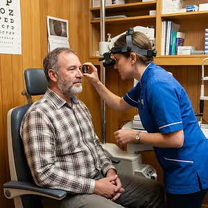
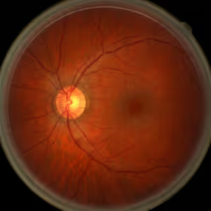
How diabetes quietly harms the retina
High blood sugar stresses the walls of the tiny vessels that feed the retina. Over time, those vessels can leak, close off, or grow in abnormal patterns — a condition known as diabetic retinopathy, and a leading cause of vision loss in working-age adults in the United States.
Because the retina has no pain receptors, early changes often develop without any noticeable symptoms. Annual evaluation gives Dr. Malani the ability to find and document changes while treatment options are broadest.
When to schedule a Diabetic Eye Exam
- A diabetes diagnosis within the past year, even with good control
- A1C that has been difficult to keep in range for several months
- Vision that shifts with blood sugar fluctuations throughout the day
- More than 12 months since your last eye exam
- Blurry central or peripheral vision that changes day to day
- New floaters, dark spots, or strands in either eye
- Other diabetes complications already confirmed by your medical team
- A new or adjusted insulin regimen that has changed your glucose patterns
What Lotus Eyecare evaluates and documents
Wide-field retinal photography captures the full extent of the retina in a single image, allowing Dr. Malani to compare each visit against prior years to track whether vessel patterns are stable or changing. OCT imaging measures fluid and swelling in the macula — the area most often affected by diabetic changes to central vision. Both tests are non-contact and completed in a few minutes.
After every diabetic eye exam, Dr. Malani sends a written report to your endocrinologist or primary care provider, describing retinal findings in clinical terms your medical team can use. Coordination between eye care and overall diabetes management is built into every visit.
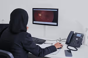
Wide-field imaging documents every retinal vessel across the entire fundus. OCT measures the precise thickness of each layer of the macula. Together they give Dr. Malani a quantitative record she can compare year to year, so progression never goes unnoticed between visits.

After every diabetic eye visit, a written clinical summary goes directly to your endocrinologist or primary care doctor. It details retinal findings, grading of any retinopathy, and whether the current diabetes management plan is protecting the eye. That coordination is built into every eye exam at Lotus Eyecare.
What happens during the visit
Plan on about an hour. Dr. Malani reviews your diabetes history, current A1C, and any symptoms before examining the front of the eye and imaging the retina. She walks you through the images, explains what is stable and what warrants monitoring, and gives you a clear return interval based on what she sees. If findings suggest a retinal specialist is needed, Lotus Eyecare has referral relationships in place to keep that next step moving without delay.
- Full vision assessment including refraction
- Intraocular pressure measurement
- Wide-field retinal photography
- OCT of the macula when indicated
- Pupil dilation when clinically warranted
- Written report sent to your medical team with consent

Other medical eye care at Lotus Eyecare
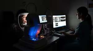

Comprehensive Eye Exam
A thorough exam checks vision and eye health, not just your prescription.

Dry Eye Treatment
Envision IPL and iLux therapy for lasting relief from evaporative dry eye.
Protect your vision — schedule your annual Diabetic Eye Exam today
Annual diabetic eye exams at Lotus Eyecare in Frisco help keep your eye health and your full medical team in sync.
Diabetic Eye Exam questions Frisco patients ask
Once a year is the standard for most adults with type 1 or type 2 diabetes. If retinopathy is already present, Dr. Malani may schedule follow-up at six months or sooner depending on severity. Newly diagnosed patients should be seen within the first few months to establish a clear baseline before any changes develop.
Usually yes. Dilation opens the pupil wide enough to see the full retina rather than just the center. Wide-field imaging captures a large field on its own, but dilation remains the clinical standard for detecting peripheral hemorrhages, neovascularization, or subtle vessel changes near the ora serrata. Plan on light sensitivity for two to four hours and bring sunglasses.
Yes. Diabetic retinopathy is a medical condition, so the exam typically bills to your medical insurance rather than a vision plan. Lotus Eyecare verifies benefits before the visit and communicates what to expect for any copay or deductible so there are no surprises at checkout.
Yes, and this is the most important point: early diabetic retinopathy produces no symptoms at all. Vision feels completely normal at the stage where treatment is most effective. The annual exam exists precisely to find damage before patients can sense it, when options are still simple and reversible.
Always, with your permission. After every diabetic eye exam, Dr. Malani sends a written clinical summary to your endocrinologist or primary care provider. The report includes retinal grading, imaging findings, and any recommendation for follow-up. That connection between eye care and systemic care is why annual exams matter beyond just checking your vision.